The Regional Burns and Paediatric Plastic Surgery Service at Alder Hey provides a service to the whole of Merseyside and Cheshire as well as North Wales and the Isle of Man. Plastic surgery involves the management and correction of congenital and acquired conditions that lead to visible differences or functional compromise. The term plastic surgery comes from the Greek work “plastikos” meaning ‘to mould’ – referring to the surgical ‘remoulding’ of physical differences.
Why would my child need plastic surgery?
One of the most common reasons children attend the department is because of an injury and our trauma service is one of the busiest in the country.
Another common reason is due to a congenital difference that may lead to difficulty with some activities or a visible disfigurement. Sensitively managed with our team approach, we aim to help a child to overcome the obstacles they may face.
Our services
We provide a general plastic surgery service to treat conditions like moles, cysts and birthmarks, correction of prominent ears and tongue-tie.
Plastic surgeons also manage soft tissue injuries from trauma – like burns, lacerations, crushed fingertips, hand fractures and dog bites, and also work on more complex cased like reattaching amputated limbs or facial reconstruction.
We offer an upper limb service, dealing with everything from extra digits to complex hand restructions, and a craniofacial service. We also have a burns service, facial palsy service and treat vascular anomalies. You can read more about these in the tabs below.
Early Bird clinic
Our early bird clinics cut waiting times for plastic surgery treatment and help children and parents spend less time spent in hospital – on average now, just half a day.
How does the Early Bird Clinic work?
- Patients are initially assessed at their local community healthcare centre
- If plastic surgery is required, patients are referred to the 7.30am Early Bird Plastic Trauma assessment clinic. Parents may be given preparation instructions e.g fasting overnight
- The patient is seen at the Early Bird clinic and if the child meets the specific eligibility criteria they are admitted for surgery immediately, with parents receiving comprehensive post-operation information
- After surgery, the patient is monitored and then discharged later the same day
As well as families having to spend much less time in hospital, the new system means young patients can prepare for surgery by fasting in their own home and then return home much sooner after their operation.
Burns services
We treat all levels of burn injury in children, including those severely injured who need intensive care.
The burns multidisciplinary team comprises experienced nurses, two consultant burns surgeons, an Associate Specialist surgeon, physiotherapists, occupational therapists, dietitian, psychologists, intensivist, anaesthetist, audit assistant and burn research nurse.
In addition to a burns outpatient service, we also have a scar management clinic, a pressure garment service and a multidisciplinary scar reconstructive clinic.
We also offer a burns support club which provides extra-curricular activities in holiday times.
Facial Palsy service
Facial palsy is a weakness in the muscles of the face. This is most commonly caused by damage to the facial nerve that provides the nerve supply to the fine muscles that are responsible for facial movement. This results in an inability to perform normal facial expressions.
This palsy can be due to damage to the nerve at any point along its course from the brain through the skull and in the face where it forms five main branches. The palsy can be complete (a total loss of function) or partial (a weakness rather than complete loss of movement) affecting all divisions of the nerve or only specific branches. As a result, there is great variability in the way that children with facial palsy are affected.
Causes of facial palsy
There is a great deal of information on the Facial Palsy UK website and we recommend you spend some time exploring it; here we outline the impact facial palsy can have and some of the numerous causes.
Laser treatment
Find more information about what to do before and throughout the course of laser treatment.
More informationMeet the surgeons
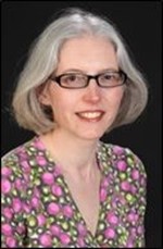
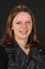
Ms Sian Falder, Consultant Plastic Surgeon
Clinical Lead of the Burn Service
MB BS, BSc, FRCSEng, FRCS(Plast), DTM&H
Sian Falder has been a consultant in burns and plastic surgery since 2008. She qualified in Medicine from St Mary’s Hospital Medical School (now part of Imperial College) in 1992 and obtained a First Class degree in the History of Medicine. She trained in plastic surgery in Bristol and Merseyside obtaining the specialist qualification in plastic surgery FRCS(Plast) in 2005. She also holds a Diploma of Tropical Medicine. Sian undertook a fellowship year of training in burns in Australia. She is the Lead Clinician for the Burn Service and is a member of the British Burn Association research special interest group and the National Burn Standards Committee. She has published a number of academic papers. She leads Alder Hey’s partnership with Kanti Children’s Hospital in Kathmandu, Nepal and works with Interburns charity. She has taken up the role of Director of Clinical Effectiveness and Service Transformation in Alder Hey and attends Trust Board.
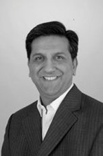
Mr Adel Y Fattah, Consultant Plastic Surgeon
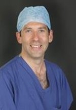
BSc(Hons), PhD, MB Bchir, FRCS(Plast)
Adel Fattah began his career at the University of Wales College of Medicine and during the early part of his training received an MRC award to study for a BSc(Hons) in Anatomy. Adel received a First Class degree and during this period investigated synovial joint development. This research was awarded the Jakoby Prize and he was subsequently recruited to the Department of Craniofacial Development at the University of London to undertake a PhD in the molecular control of joint development and craniofacial development.
After a brief post-doctoral period, he returned to complete his medical training at the University of Cambridge. His post-graduate training was undertaken in London and included terms at Great Ormond Hospital and the St Andrew’s Centre for Burns and Plastic Surgery, where he gained good experience in burns care. He was awarded the FRCS(Plast) in 2009 and was entered onto the Specialist Register for Plastic Surgery in 2012 following fellowships abroad at “SickKids” in Toronto, the largest paediatric hospital in North America. Adel has maintained active research interests since his PhD and has won a number of prizes for his work presented at over 40 national and international conferences. He has also published over 35 articles mainly regarding facial palsy, craniofacial reconstruction and microsurgery. He also acts as a reviewer for a number of academic journals. Adel is an avid teacher and has produced teaching videos for a number of surgical procedures and is committed to training the next generation of plastic surgeons.
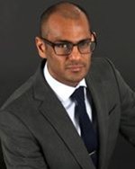
Mr Christian Duncan, Consultant Plastic Surgeon
CBU Lead (Neuroscience, Musculoskeletal & Specialist Surgery)
MB BCh, BAO, MPhil, FRCSI, FRCS(Plast)
Mr Duncan graduated as a doctor in 1991 from University College Dublin and undertook basic surgical training in Dublin, gaining his general fellowship in 1995. He undertook higher surgical training in plastic surgery in Bristol, Sheffield and the West Midlands gaining FRCS(Plast) in 2003. During higher surgical training, he pursued fellowships in Tissue Engineering at the School of Dentistry in Birmingham, Craniofacial Surgery in the West Midlands Supra-Regional Craniofacial Unit and Aesthetic Surgery at the Wellington Hospital, London. He was awarded an MPhil for research in tissue engineering of skin in 2002. He was appointed as a Consultant Plastic Surgeon at Alder Hey NHS Foundation Trust in 2004. He joined the Merseyside Supra-Regional Craniofacial Team in 2005 and became the clinical lead of that team in 2010. Clinical interests include all aspects of craniofacial surgery and research interests include tissue engineering, cell delivery systems and bone cements. His research has been widely published internationally.
Mrs Catherine Raraty, Associate Specialist Plastic Surgeon
MB ChB, FRCSEd MmedSci
Mrs Raraty qualified as a doctor from Leeds in 1989 and became a Fellow of the Royal College of Surgeons in 1997. She first worked in the plastic surgery department at Alder Hey in 1992 and came back many times during training. She returned to Alder Hey full time in 2003 as a Fellow, becoming a Specialty Doctor in 2007 and an Associate Specialist in 2009. Her main responsibility is the care of burn inpatients and their clinical follow up after discharge. She is part of the Clinical Advisory Group for the Northern Burn Care Network and a Key Instructor for the Emergency Management of Severe Burns Course (EMSB). Mrs Raraty also carries out general plastic surgery lists and clinics. She is the Clinical Governance Lead for the Burns & Plastic Surgery Department.
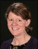
Ms Eleonore Breuning, Consultant Plastic Surgeon
MBBCh (UWCM 2000), MD (Birmingham 2013), FRCS(Plast) (London 2012)
Eleonore Breuning started in Alder Hey in January 2016, having completed a one-year Fellowship in Paediatric Plastic Surgery in British Columbia Children’s Hospital in Vancouver in 2014/15. She qualified as a doctor in 2000 from the University of Wales College of Medicine and then completed basic surgical and specialist plastic surgery training in both adult and paediatric plastic surgery in the West Midlands region. She undertook an 11 month Hand Surgery fellowship in Birmingham in 2014 and gained additional experience in burns care. Her clinical interests include Vascular Anomalies, Hand Surgery and Lasers, including improvement of burn scarring. Her research interests include Patient Reported Outcome Measures (PROMs). Specialist areas are general paediatric plastic surgery, lasers and vascular anomalies, hand surgery.
Mr Zahid Hassan, Consultant Plastic & Hand Surgeon
BM, MRCS, MD, FRCS(Plast), Dip Hand Surg (Br)
Zahid Hassan graduated from Southampton University in 1996. Basic surgical training was completed in New Zealand, London and Swansea leading to the qualification Member of the Royal College of Surgeons of England (MRCS) which was gained in 2001. At Manchester University he was awarded a higher doctorate (MD) for research into paediatric burn wound healing. Higher plastic surgical training was undertaken in the Mersey region leading to the Intercollegiate Specialty Fellowship in Plastic Surgery FRCS(Plast) in 2012. He subsequently completed the prestigious Advanced Training Fellowship in Hand Surgery at Leeds for 12 months. During this period he also obtained the British Diploma in Hand Surgery. He was appointed as a Consultant in Plastic, Reconstructive and Hand Surgery at Whiston Hospital in 2014 and joined the team at Alder Hey Hospital in 2015. Mr Hassan is qualified in the Emergency Management of Severe Burns. Specialist areas include congenital hand abnormalities, nerve reconstruction, cerebral palsy and upper limb trauma.
Mr Pundrique Sharma
BSc(Hons) PhD MBBS FRSC(Plast)
Mr Sharma has a special interest in paediatric microsurgery, limb reconstruction and nerve injury. He attended University College London (UCL), and was selected against national competition to be one of only a six students to do a combined medical degree and PhD. He qualified as a doctor in 2003 and spent his junior years training in London. Pundrique undertook specialist training in plastic surgery in and around London and the South East, including at UCL, the Cambridge University Hospitals NHS Trust, and the St Andrew’s Centre for Plastic Surgery and Burns in Chelmsford. After attaining FRCS(Plast) exam, he had further training in head and neck reconstruction and microsurgery at the Norfolk and Norwich University Hospital, and in reconstructive microsurgery at Chelmsford. He also undertook sub-speciality fellowships in at Great Ormond Street Hospital and Sick Kids Hospital in Toronto, training in all aspects of paediatric plastic surgery but in particular focussing on limb and facial reconstruction.
Physiotherapy: Helen Hartley and Rebecca Pratt are our therapists that employ a range of techniques to treat some of the symptoms of facial palsy such a facial spasm as synkinesis as well as having a key role in post-operative rehabilitation following smile reconstruction.
Psychology: Rachel Mumford and her team provide psychological support to those children and families that experience difficulties coping with facial palsy.
Speech and Language Therapy: Wendy Blumenow is also a member of the craniofacial service and provides speech therapy to the small number of patients that have issues in this area.
Neurology: Dr Ram Kumar and Dr Stefan Spinty are the neurologists on the team who is involved in diagnosis and treatment of facial palsy. Dr Kumar graduated in medicine at University of Cambridge in 1995. He has been a consultant paediatric neurologist at Alder Hey since 2007. Dr Kumar has contributed chapters to a number of textbooks and is actively engaged in research, undergraduate and postgraduate multiprofessional education.
Neurosurgery: Mr Conor Malluci is our neurosurgeon that has a practice in brain tumours; some patients will experience a facial palsy following tumour removal and Mr Malluci is our link for children that present with facial palsy that is found to be due to a tumour. He has just finished his tenure as chairman of the British Paediatric Neurosurgery Group and continues to chair their research group. He is currently deputy editor of the British Journal of Neurosurgery and the associate editor of the European journal, “Child’s Nervous System”.
Ear Nose and Throat: Mr Ravi Sharma is our ENT surgeon.
Plastic surgery: We have a dedicated ward for our facial palsy patients with individual cubicles and en suite facilities for families. Mr Adel Y Fattah is the lead for the facial palsy service having trained in Toronto with Dr Ron Zuker. Maria Kelly is our specialist nurse and is responsible for liaising between specialties in order to coordinate appointments.
Theatre Team: We have a dedicated core group of theatre staff that perform our facial reanimation and microsurgery cases.
How to refer
Referrals will be accepted via the Regional Paediatric Burns and Plastic Surgery Service and the Choose and Book system.
Email: [email protected]
Choose and Book: Please search under Alder Hey Plastic Surgery
Address:
Facial Nerve Programme
Regional Paediatric Burns and Plastic Surgery Service
Alder Hey NHS Children’s Foundation Trust
Eaton Road
Liverpool
L12 2AP